Evaluation of the Anticancer Property of Makahiya (Mimosa Pudica L.) and Paper Flower (Bougainvillea Glabra Choisy) In Inhibiting Tumor Angiogenesis
Bacan, Cleford Jay D.
Ellaga, Mark Jobert C.
Facal, Joygie Nessa R.
Hermosilla, Keith Emmanuelle H.
Inoue, Shiori S.
Lanit, Ace Bernard H.
Lausa, Vicelda Karla G.
Quidet, Princess Lourdes C.
Rabadon, Alexandra A.
Rojo, Althea Shane C.
Cor Jesu College, Inc. – Basic Education Department , Digos, Davao Del Sur, Philippines
Over time, cancer has remained widespread, causing profound harm to patients due to its numerous forms. The disease arises when healthy cells transform into malignant tumors. The commonly employed methods for combating cancer include surgery, radiotherapy, and chemotherapy. Nevertheless, these treatments have enduring and harmful impacts on the body, such as disrupting the growth of healthy cells, subjecting patients to radiation, and potentially affecting the organs in the treated region. Furthermore, the current treatments have overlooked the potential of utilizing the inherent anticancer properties found naturally in plants. Thus, makahiya and Paper flower were found to possess phytochemicals, which have the potential to inhibit angiogenesis. To evaluate this potential, the study aimed to assess their anti-angiogenic properties using the chorioallantoic membrane (CAM) assay. For this purpose, thirty fertilized duck eggs were obtained and used as test subjects in the experimental procedure. The study which was conducted over a period of three months from February to April, collected the data through the software “ImageJ” to obtain the blood vessel measurements of the eggs. The researchers used the analysis of variance (ANOVA) to analyze the data. Then, the post hoc comparisons using the Tukey’s HSD test were used to compare the results of the four treatments to test the hypothesis of the effectiveness of the makahiya and Paper flower extracts. The results show the significant differences of the treatments in comparison to the control group. Moreover, the statistics indicated both having significant and no significant differences in measurements before and after the treatment, respectively. Therefore, the extracts from both plants are feasible for angiogenesis inhibition.
Keywords: angiogenesis, makahiya, paper flower, CAM assay, ImageJ
Cancer has been prevalent throughout the years, with its countless types leaving devastating effects on patients. Its development is the result of healthy cells turning into malignant tumor cells. The most well-known procedures in dealing with cancer are surgery, radiotherapy, and chemotherapy (Debella et al., 2021). However, those procedures leave lasting and harmful effects on the body, such as affecting the growth of healthy cells and exposure to radiation, and may affect the organs of the treated area (Amjad et al., 2023). In addition, there has been a lack of using naturally occurring alkaloids and flavonoids in plants in the currently available treatments.
The World Health Organization (2022) reported that cancer has claimed around 10 million lives in 2020. Lung cancer is recognized as the second most commonly diagnosed cancer and the primary contributor to cancer-related deaths in the United States (Schabath & Cote, 2019). According to the American Cancer Society (2018) and Bray (2018), globally, lung cancer has consistently held the title of the most prevalent cancer, reporting 2.1 million new cases in 2018, which accounted for 12% of the overall global cancer burden. Among its various forms, breast cancer emerges as the most prevalent, affecting approximately 2.3 million women worldwide and leading to 685,000 deaths. In terms of leading types of cancer, lung, tracheal, and bronchus cancer ranked as the top contributor to cancer-related fatalities in both males and females, with colon and rectum cancer, esophageal cancer, and stomach cancer being major causes in males (Tran, 2019).
Moreover, the treatment of cancer places a substantial strain on medical resources, and the morbidity and mortality associated with the disease create a significant societal burden, making cancer a crucial economic challenge across the globe (Wang et al., 2023). Different types of cancers have not only claimed lives but have continued to stand as a formidable global health challenge while being an economic burden, thus posing a greater need to control the disease.
Over 1 million new cases of cancer and over 700,000 cancer-related deaths occurred in the ASEAN region in 2020. Among these cases, breast, lung, colon, and rectum cancers were the most frequently diagnosed types of cancer. In contrast, lung, liver, and breast cancers were the leading causes of cancer-related fatalities (Alberto et al., 2023). Since 2000, cancer has been Thailand’s primary cause of death, with rates of malignant neoplasms continuously rising (Insamran & Sangrajrang, 2020). Moreover, cancer rates are rising in Indonesia, a middle-income nation with over 270 million people. According to the Global Cancer Observatory (2020), there were 141.1 new instances of cancer per 100,000 people and 85.1 cancer deaths per 100,000 people (Puspitaningtyas et al., 2021). In 2019, the World Health Government indicated that there were around 115,000 cases of cancer-related deaths and 165,000 new cancer cases in Vietnam. Vietnam is ranked 57th internationally with 104 per 100,000 people age-standardized cancer death rate. These statistics highlight the urgent need for more comprehensive strategies addressing prevention and effective treatment to combat the growing cancer burden in the ASEAN region.
Furthermore, in the Philippines, cancer has emerged as the second most common cause of death, driven mainly by breast, lung, colorectal, liver, and prostate cancers (Trinidad, 2019). The World Health Government (2021) stated that there are over 4700 annual childhood cancer diagnoses, primarily leukemia and other types of cancer affecting various organs, leading to approximately 1700 deaths each year with an estimated population of 107 million in the Philippines.
As well as in the local context, the number of cancer patients in the Southern Philippine Medical Center-Adult Cancer Center (SPMC-ACC) increased to 4,470 patients in 2022 compared to the previous year (Patumbon, 2023). Moreover, Dr. Aldrich Kyne So, an SPMC Level II resident doctor, mentioned in an interview that the Philippines’ most common cancers include breast, cervical, colorectal, prostate, and lung cancer. However, most cancer patients in Davao City experience breast cancer (Oblianda, 2023). As the community struggles with these health challenges, there is an urgency to develop an intervention to address the prevalence of cancer in the region.
Cancer is a rampant disease, leaving a lasting effect on patients from different nations turning healthy cells into malignant tumor cells. Given its nature, there is a lack of safer procedures for alleviating the spread of tumor cells than the commercially available treatments, such as radiotherapy and chemotherapy, which can expose nearby organs to radiation in the treated area (Willamette Valley Cancer Institute and Research Center, 2023). Moreover, there is a need for more exploration into the anti-angiogenesis properties of plants, such as makahiya and paper flowers, as most studies only address their phytochemical properties and lack the testing of those properties. This study aimed to make use of the unutilized makahiya (Mimosa pudica L.) and Paper flower (Bougainvillea glabra Choisy) as components in inhibiting tumor cell division. The researchers sought to discover the efficacy of anticancer properties such as phytochemicals, flavonoids, and alkaloids in inhibiting tumor angiogenesis.
Statement of the Problem
The main objective of this research is to test the efficacy of Makahiya (Mimosa pudica L.) and Paper flower (Bougainvillea glabra Choisy) in alleviating the division of tumor cells. Specifically, the study sought to answer the following questions:
What is the efficacy of using Makahiya (Mimosa pudica L.) in alleviating cell division in terms of:
Vascularity count before plant extract solution; and
Vascularity count after administration of different concentrations of plant extract?
What is the efficacy of using Paper flower (Bougainvillea glabra Choisy) in alleviating cell division in terms of:
Vascularity count before plant extract solution; and
Vascularity count after administration of different concentrations of plant extract?
What is the efficacy of using Makahiya (Mimosa pudica L.) and Paper flower (Bougainvillea glabra Choisy) in alleviating cell division in terms of:
Vascularity count before plant extract solution; and
Vascularity count after administration of different concentrations of plant extract?
Is there a significant difference in alleviating the division of tumor cells using the anticancer properties of the Makahiya (Mimosa pudica L.) and Paper flower (Bougainvillea glabra Choisy) plant?
Hypothesis
To answer the problem listed in the preceding section objectively, the given null hypothesis was formulated:
H0: There is no significant difference in alleviating the division of tumor cells using the anticancer properties of the Makahiya (Mimosa pudica L.) and Paper flower (Bougainvillea glabra Choisy) plant.
Significance of the Study
It is critical to explore the alkaloidal property of Makahiya (Mimosa pudica L.) plant and Paper flower (Bougainvillea glabra Choisy) extract in inhibiting tumor cell division as it will contribute to understanding of its potential benefits in tumor inhibition. Hence, the researchers conducted this study, which could be beneficial to the following:
Department of Health. This study can benefit the Department of Health for several reasons. In acquiring profound knowledge of the alkaloidal properties of the Makahiya Paper flower in their potential to alleviate tumor cell division through this quantitative study, healthcare professionals of the Department of Health can use the data and information of this research. It can potentially impact or contribute to cancer prevention, treatment effectiveness, patient outcomes, and overall progress in the fight against cancer.
Cancer Centers. This study can significantly benefit cancer centers from its potential findings as it may revolutionize cancer centers by providing innovative insights, enhancing treatment strategies, improving patient outcomes, and advancing the overall understanding of cancer biology, thus elevating the standard of care and fostering hope for patients and their families. Moreover, the potential impact of these groundbreaking findings extends to advancing the development of more targeted approaches in cancer treatment.
Future Researchers. This study holds great potential for future researchers, as it aims to provide comprehensive knowledge regarding the alkaloidal properties of the Makahiya and Paper flower plants and its potential in mitigating tumor cell division. The findings of this study can serve as a valuable reference and guide for the future development of related research endeavors, shedding light on new possibilities and avenues of investigation.
Scope and Limitations
This study focused mainly on investigating the anti-cancer properties of Makahiya (Mimosa pudica L.) and Paper flower (Bougainvillea glabra Choisy) extract in alleviating tumor cell division and its efficacy. The research was conducted during the first and second semesters of the academic year 2023 to 2024. This research was conducted in Digos City, Davao del Sur. Moreover, for the research testing, the researchers utilized a duck embryo within the province of Davao del Sur. This study did not specify a particular tumor for investigation; instead, it focused on examining the anti-cancer properties of Makahiya (Mimosa pudica L.) and Paper flower (Bougainvillea glabra Choisy) extract in inhibiting tumor cell division and assessing their efficacy.
Definition of Terms
The following terms were expounded to further understand the components of this study.
Alkaloidal Property. This refers to the characteristic or quality of having alkaloids obtained from the Makahiya and Paper flower extracts. Alkaloids are naturally occurring organic compounds that frequently have pharmacological effects. The alkaloidal properties specifically pertain to the alkaloids extracted from the makahiya and Paper flower plants, which may exhibit various biological activities, including potential anti-cancer properties.
Angiogenesis. This refers to the process of creating new blood vessels from existing ones, which is crucial for the growth and spread of tumors. Tumors rely on a sufficient blood supply to obtain the necessary nutrients and oxygen for continuous growth.
Antiangiogenesis. This term refers to the deliberate process of inhibiting the new blood vessel formation in the embryo’s growth. This process is designed to impede the development of blood vessels that would otherwise provide essential nutrients and oxygen to the embryonic tumor model. This inhibition aims to curtail the embryo’s growth with tumor-like characteristics and prevent the potential spread of these simulated tumor cells.
Blood vessels. This refers to one of the key aspects of the study, as they are the channels through which blood is distributed in the embryo. These vessels will be monitored to observe its number of growths throughout the experiment.
Cell Division. This refers to the process in which the alkaloids found in the Makahiya (Mimosa pudica L.) plant and Paper flower (Bougainvillea glabra Choisy) plant will inhibit or slow down, specifically the process of tumor cell division, potentially providing a basis for anti-cancer treatment.
Concentration. This is the amount of makahiya and Paper flower extracts that will be used in the experiment. The researchers will test different concentrations to determine the plant extracts’ effectiveness.
Flavonoid. It is a common class of phytochemicals that possess antimicrobial, antioxidant, anticancer, anti-inflammatory, and wound-healing characteristics.
Makahiya plant. This refers to the extract that has potential phytochemical properties and their impact on inhibiting tumor cell division.
Paper Flower. This refers to the extract possessing potential phytochemical properties and its ability to impact the inhibition of tumor cell division.
Tumor Cells. This refers to the egg embryo model that exhibits characteristics similar to tumor cells, aiming to investigate how the potential anticancer properties of the Makahiya (Mimosa pudica L.) plant and Paper flower (Bougainvillea glabra Choisy) may affect the division and growth of these cells within the embryo model.
Makahiya Plant (Mimosa pudica L.)
The makahiya plant, known for its sensitive leaves, holds promising potential in traditional medicine due to its diverse medicinal properties and therapeutic applications. Plants historically used for medicinal purposes contribute significantly to drug development, with 75% of market drugs derived from them (Jagetia & Vanlalhruaii, 2020). Makahiya is a common and abundant plant in the Philippines, known for its resiliency and adaptability (Cruz, 2021). It belongs to the Fabaceae family and is a well-known traditional plant containing several beneficial secondary metabolites, including triterpenes, tannins, steroids, flavonoids, and glycosyl flavones. The plant as a whole is precious for a variety of pharmacological and biological activities. Its plant components possess various pharmacological properties, including antioxidant, antifungal, anti-inflammatory, antibacterial, and wound-healing activity (Fernandes & Santosh, 2022; Rajendiran et al., 2022; Rathnamali, 2019).
Furthermore, makahiya plants are traditionally utilized for urogenital issues, piles, dysentery, sinus conditions, and topical wound treatments. It has been employed for the prevention and treatment of several disorders, such as cancer, diabetes, hepatitis, obesity, and urinary infections (Rathnamali, 2019).
The makahiya plant is also known for its abundance of primary and secondary metabolites, which contribute to their pharmacological potential. A phytochemical analysis revealed the presence of 40 recognized chemical components, primarily alkaloids, phenols, and flavonoids (Bole et al., 2021; Kumar, 2020; Rizwan et al., 2022). It is often found in the leaves, flowers, and roots (Das et al., 2021). Rathnamali (2019) also claims that the roots and leaves of makahiya have exhibited the most significant pharmacological activity. In the study of Jagetia (2018), those compounds were identified as the reason for their traditional use in the Hindu system of medicine in treating different diseases. Similarly, the findings of Mandal et al. (2022) suggested that makahiya is a promising plant with therapeutic potential due to the presence of different phytochemicals such as flavonoids, saponins, coumarins, tannins, terpenoids, and alkaloids.
Furthermore, makahiya plant was found to be a valuable natural source of antioxidants (Jagetia, 2018). Additionally, Braganza (2023) harbors specific antioxidants that have the potential to counteract detrimental free radicals within the body, possibly aiding in the enhancement of overall health.
Recent studies have highlighted the plant’s alkaloid content, particularly mimosine, which possesses anticancer properties, further enhancing the plant’s reputation. A chemical analysis revealed the presence of alkaloids in all the extracts of Jungle geranium (Ixora coccinea) and Makahiya (Mimosa pudica L.) (Cua et al., 2018). When the alkaloid content was measured and compared to the in vitro anti-acid test results, it was found that there was a positive relationship between alkaloid content and its buffering capacity activity. Another study by Tripathi and Soni (2022) discovered that its roots and leaves exhibit significant effects against diabetes, oxidation, liver damage, antioxidants, wound healing, diuretics, cancer, and seizures.
A study conducted by Masagca et al. (2020) on makahiya highlighted its examination for anti-angiogenic effects. The study demonstrated that the aqueous root extract of makahiya effectively suppressed angiogenesis. Extracts from Mimosa, particularly in water and ethanol, exhibit potential toxic effects on Dalton’s Ascitic Lymphoma cell, possibly through triggering membrane damage, lipid peroxidation, and increased lactate dehydrogenase levels, leading to a nonapoptotic form of cell death. The varying activity levels among extracts are attributed to differences in their secondary metabolite composition (Jagetia & Vanlalhruaii, 2020).
Paper flower (Bougainvillea glabra Choisy)
Paper flower (Bougainvillea glabra Choisy) is renowned in traditional medicine for treating respiratory issues like cough, asthma, bronchitis, and gastrointestinal diseases. Its effectiveness extends to antibacterial and insecticidal properties. It contains several biochemicals, such as alkaloids, flavonoids, betacyanin, and pinitol. It was also discovered that Paper flowers contain various compounds, including glycosides, anthraquinone, terpenoids, saponin, fat, and tannin. An analysis confirmed the existence of alkaloids, flavonoids, phenolic compounds, and tannins by applying distinct qualitative tests tailored for detecting these specific phytochemicals (Gupta & Gupta, 2021).
Despite the rich presence of secondary metabolites like alkanes, phenols, terpenes, and betalains in the plant’s involucre, its antimicrobial potential remains unexplored (Garcia et al., 2023). A recent study by Hanggag and Elhaw (2022) uncovered the phytochemical content in different parts, focusing on leaves, bracts, and flowers. The study revealed that bracts contained the highest level of active minerals among the various plant parts.
The pharmacological properties of Paper flower extend to other species. The article of Anurag et al. (2022) covers an array of Paper flower species, cultivars, and hybrids, exploring their medicinal, pharmacological, and toxicological uses. While the pharmacological potential of Paper Flower remains partially investigated, evidence suggests that this plant genus exhibits anti-inflammatory, antioxidant, immune-modulatory, antibacterial, and other advantageous effects in animal studies. Paper flower plays a crucial role as a rich source of medicinal compounds with diverse properties such as anti-diabetic, antispasmodic, emetic, anti-cancer, antimicrobial, anti-inflammatory, insecticidal, anti-ulcer, antimicrobial, anti-diarrheal, and antiviral effects. The extracts exhibited significant biological effects linked to elevated levels of bioactive compounds, unveiling the potential utilization of Paper flowers as a natural source of potent bioactive agents, including antioxidants, enzyme inhibitors, and potential anticancer agents, laying a new foundation for their application (Kalaiyarasan et al., 2022; Saleem et al., 2019; Teh et al., 2019).
In a separate study by Saleem et al. (2021), several authors have suggested the potential of Paper flower as a therapeutic option for various cancers. However, elucidating its precise molecular targets necessitates rigorous scientific methodologies like apoptosis and flow cytometry. Limited in-vivo animal model-based research has been conducted, revealing limitations in establishing controls and performing dose-effect analyses in antimicrobial, antioxidant, and antidiabetic investigations.
The Paper flower and Madagascar Periwinkle (Catharanthus roseus) plant extracts underwent various qualitative chemical tests to explore their chemical composition. The investigation of the study confirmed the presence of alkaloids, flavonoids, phenolic compounds, and tannins in the extracts (Tegeli, 2019). The methanol extract is abundant in bioactive compounds, possesses significant antioxidant properties, and can inhibit enzymes. Both extracts displayed varying levels of cytotoxicity against breast, cervical, prostate, and colon cancers. Ultra-High Performance Liquid Chromatography-Mass Spectrometry (UHPLC-MS) analysis indicated the potential presence of 27 secondary metabolites, predominantly terpenoid, alkaloid, and phenolic derivatives.
Regarding enzyme inhibition, all the extracts displayed moderate effectiveness against tyrosinase and α-amylases. However, regarding liquid oxygen, the methanolic extracts exhibited superior percentage inhibition compared to the Dilated Cardiomyopathy extracts. These findings suggest that Paper flower holds promise as a foundational resource for developing innovative pharmaceuticals (Saleem et al., 2020).
Various phytochemical compounds found in methanolic extracts of Paper flower leaves can effectively shield the body against oxidative damage caused by free radicals. Free radical-mediated DNA damage can cause cell mutagenesis and initiate cancer development. Paper flower aerial dichloromethane extract is effective against breast cancer and cervical cancer. Paper flower aerial methanol extract has demonstrated anti-proliferative solid activity for colon cancer cell lines (Information Resources Management Association USA, 2022).
Anticancer Properties
Extensive research and empirical data highlight flavonoids’ diverse potential and multifaceted roles in cancer prevention and management. Phytochemicals found in plants, including taxol variations, vinca alkaloids (vincristine, vinblastine), and podophyllotoxin analogs, exert influence on cancer pathways by immune regulation, cell cycle arrest and the suppression of proliferation (Choudhari et al., 2020). Furthermore, they play a pivotal role in chemoprevention by safeguarding against DNA damage and repairing mutated genes, offering a viable cancer prevention strategy through dietary adjustments (Surampudi, 2023). Gaikwad and Srivastava (2022) extensively explored the anti-cancer potential of phytochemicals in vitro and in vivo, advocating their suitability as anti-cancer medicines owing to their safety profile and well-researched mode of action.
Moreover, a direct link was established between phytochemical intake and health benefits, advocating the consumption of foods rich in phytochemicals such as carotenoids, polyphenols, and isoprenoids for disease prevention. The effectiveness of these compounds is contingent upon various factors such as extraction methods, solvents, temperature, and time (Kumar et al., 2023). Building upon this understanding, Soni and Duddukuri (2023) delved deeper into the molecular mechanisms of plant extracts and phytochemicals, highlighting their diverse targets in cancer progression. The empirical data underscores the chemo-preventive characteristics of phytochemicals and their efficacy against cancer-promoting agents. Furthermore, Javed (2020) emphasizes the significance of medicinal plants as abundant sources of compounds like artemisinin, vinblastine, and vincristine, contributing significantly to drug development. Recent advancements in extraction and screening techniques from natural sources have yielded substantial pharmaceutical discoveries.
Investigating natural alkaloids uncovers their remarkable capacity to impact diverse mechanistic pathways associated with cell proliferation, regulation of the cell cycle, and metastasis. This comprehensive review highlights alkaloids’ potential as agents for anti-colon cancer chemotherapy, demonstrating their capacity to modulate or halt the cell cycle (Khan et al., 2022; Olofinsan et al., 2023). Preclinical research demonstrates the ability of these alkaloids to inhibit the growth of colon cancer cells by causing cell cycle arrest at various stages. This suggests their potential as highly effective anticancer agents with promising prospects. Additionally, a thorough examination of naturally occurring alkaloids known for their anticancer properties unveils their molecular mechanisms, explores synthetic derivatives, and outlines their pharmacological profile, reviewing the significance of alkaloids as a crucial category of plant-derived anticancer drugs, signifying substantial potential for future advancements in cancer therapy (Mondal et al., 2019).
Kurek (2019) emphasizes the diverse biological impacts of alkaloids present in common foods and beverages, with actions including anti-inflammatory, anticancer, analgesic, and more. Efferth and Oesch (2021) note their potential in cancer treatment, inhibiting tumor growth with effects like cell cycle arrest and apoptosis. Sadowska-Bartosz and Bartosz (2021) explore betalains, highlighting their antioxidant capabilities as potent electron donors with a phenolic compound and cyclic amine structure.
According to Bisol et al. (2019), flavonoids are recognized for their toxicity towards cancer cells while preserving healthy ones, and they are considered potential candidates for chemotherapy. Their systematic review aimed to assess flavonoids’ effectiveness in human cancer chemotherapy. With their myriad beneficial properties—from antioxidant to anticarcinogenic—as illustrated in numerous experimental and epidemiological investigations, flavonoids engage with and regulate various cellular proteins, transcription factors, and signaling enzymes. This extensive research underscores the role of flavonoids that exhibit anti-metastatic properties by interfering with tumor cell adhesion and migration as preventive and therapeutic agents for a broad spectrum of diseases, including cancer (George et al., 2021; Gurler, 2020).
Furthermore, Ullah et al. (2020) identified several classes of flavonoids, highlighting their diverse biological effects, from anticancer to cardio-protective properties. The researchers strongly recommend incorporating diverse types of flavonoids into daily diets to maintain health and mitigate the risk of severe ailments like diabetes and cancer. Kopustinskiene et al. (2020) emphasized the diverse effects of flavonoids against cancer, proposing their potential in novel preventive strategies and highlighting their role in maintaining gut health. These findings reflect Farhan’s assertion (2023) that, despite established benefits like anticancer and anti-inflammatory properties, additional research is crucial to comprehend the intricate functions of flavonoids and identify new therapeutic approaches.
Kashyap et al. (2019) summarized the various cellular and molecular mechanisms through which flavonoids exhibit anticancer properties, including apoptosis induction, cell cycle arrest, and anti-inflammatory effects. Paliwal et al. (2021) emphasized the cost-effectiveness and safety of plant-derived compounds, particularly polyphenols and flavonoids, in cancer prevention and treatment. Similarly, Hazafa et al. (2020) highlight polyphenols’ efficiency in cancer mitigation, including flavonoids, underlining their diverse roles in prediction and management. Moreover, Asao and Asaduzzaman (2018) underscore the cancer-combating abilities of prominent phytochemicals, including anthocyanins, flavonoids, and carotenoids. Sak (2019) suggested flavonoids, such as soy isoflavones and quercetin, as potential enhancers of radiotherapy against prevalent cancers. This collective evidence underscores flavonoids’ diverse potential and multifaceted roles in cancer prevention and management.
Angiogenesis
Angiogenesis, a biological process in which new blood vessels are formed from existing ones, is critically involved in multiple biological processes and serves as a significant contributor to both normal development and the advancement of diseases. As Al-Ostoot et al. (2021) highlighted, angiogenesis’s role in cancer progression is crucial for supplying tumors with oxygen, nutrients, and growth factors, aiding their spread to distant sites. Inhibiting angiogenesis is pivotal for preventing solid tumors by limiting blood flow within specific tumor regions, leading to extensive oxygen deprivation and tissue death. Moreover, the importance of tumor angiogenesis was emphasized, involving stages like blood vessel sprouting, cell proliferation, remodeling, and regression, guided by well-documented molecular factors (Liu et al., 2023; Zuazo-Gaztelu & Casanovas, 2019).
An imbalance favoring proangiogenic molecules activates previously dormant blood vessels, fostering the angiogenic state in tumor development. Additionally, Chojnacka and Lewandowska (2019) note the potential of polyphenol-rich extracts to hinder angiogenesis by influencing critical factors and cellular mechanisms, restraining blood vessel formation, and limiting cancer cell migration and invasion, thus reducing tumor growth. Cumulative evidence from in vitro and in vivo studies supports the potential use of natural polyphenols as therapeutic agents in anti-angiogenic therapies, promising for cancer treatment.
Therapeutic inhibition of tumor angiogenesis emerges as a promising strategy, showing success in animal models and providing an alternative for treating multidrug-resistant tumors (Barberaki & Kintzios, 2019). These therapies target blood vessel growth with minimal side effects due to drug accessibility and tumor cells’ reliance on a single capillary. Nevertheless, their efficacy remains confined to immature angiogenic subsets within tumors. Notably, flavonoids act as angiogenesis inhibitors, vital for average tissue growth but detrimental in tumors due to increased blood vessel formation supporting cancer cell survival and proliferation (Forni et al., 2021).
Munir et al. (2020) investigated anti-angiogenic therapies and their impact on tumor cell development concerning phytochemicals and angiogenesis inhibition. These therapies show promise in reducing tumor cells’ drug resistance. Studies conducted on cell lines and animal models have shown that biologically active phytochemicals derived from plants and diets possess antioxidant, anti-inflammatory, anti-proliferative, and anti-angiogenic properties. Ribeiro et al. (2018) highlight the limited use of synthetic antiangiogenic drugs due to toxicity, prompting ongoing research for alternatives. Natural phytochemicals from plants or micro fungi show significant potential in treating angiogenesis-related conditions with lower toxicity than synthetics. This directs attention towards exploring natural compounds as angiogenesis modulators, potentially offering new therapies with reduced drug reliance and associated side effects.
Varghese et al. (2020) proposed using phytochemicals to target miRNAs, potentially suppressing tumor angiogenesis and inhibiting tumor growth, especially in angiogenesis-dependent cancers. Plant-derived natural compounds, influencing epigenetic factors, emerge as promising cancer prevention and treatment options. This research hints at the potential of natural compounds in effectively managing tumors while mitigating drug-related concerns.
In the research study of Shahik et al. (2021), the vascular endothelial growth factor (VEGF) is highlighted. According to the statement, VEGF is present in numerous cancer cells and has a vital function in stimulating the growth, survival, and proliferation of vascular endothelial cells. It also facilitates angiogenesis through the signaling of VEGF and the vascular endothelial growth factor receptor (VEGFR). The study investigated the potential of various alkaloid molecules as inhibitors of VEGF and identified three alkaloid candidates that showed promising results in inhibiting VEGF and VEGFR-mediated angiogenesis. Those alkaloid compounds demonstrated strong binding ability, as indicated by their high scores. As a result, the study’s findings support the idea that these alkaloids could be further developed as lead compounds to design new and effective anticancer drugs.
Based on the study of Kopustinskienė et al. (2022), it is highlighted that various plants contain flavonoids, polyphenolic substances categorized into six groups: isoflavonoids, flavanones, flavanols, flavonols, flavonoids, and anthocyanins. These flavonoids are commonly found in fruits, vegetables, and plant-based beverages such as wine, green tea, and cocoa. Known for their diverse anticancer properties, flavonoids impact enzymes scavenging reactive oxygen species (ROS), contribute to cell cycle arrest, induce autophagy and apoptosis, and limit the growth and invasiveness of cancer cells. The book by Subbaraj et al. (2021) underscores that flavonoids effectively inhibit angiogenesis and metastasis by modulating various signaling pathways. Recent research positions flavonoids as potent antiangiogenic agents because they can block these crucial pathways.
Related Studies
The rise of cancer has gained prominence globally, leading to the development and utilization of chemically-based medications to combat diverse cancer types. According to Swamy et al. (2018), existing cancer treatments have drawbacks due to their harmful impact on human health. This drives the need for alternative therapies utilizing natural plant anticancer agents. Research on polyphenols, flavonoids, and brassinosteroids highlights their potential as anticancer compounds, displaying antioxidant effects, inhibiting cancer cell growth, inducing apoptosis, targeting specific cells, and demonstrating cytotoxicity against cancer cells.
According to the book chapter authored by Tan et al. (2018), researchers propose that plant compounds hold the potential to offer beneficial effects against various types of cancers, such as cervical, colon, skin melanoma, breast, prostate, and leukemia. The ability to prevent cancer is attributed to specific molecules present in plants that possess anticancer properties. These molecules include vinca alkaloids, similar compounds, curcumin, colchicine, epigallocatechin-3-gallate (EGCG), betulinic acid, and derivatives of podophyllotoxin. Scientists have isolated these compounds from plants and made modifications to enhance their solubility, toxicity, and effectiveness.
Furthermore, in studying different aspects of cancer, such as angiogenesis, tumor growth, immune evasion, metastasis, and drug resistance, the chorioallantoic-membrane (CAM) model is a valuable tool for studying different aspects of cancer, such as angiogenesis, tumor growth, immune evasion, metastasis, and drug resistance (Fischer et al., 2022). In the study of Lokman et al. (2020), the extraembryonic membrane, or CAM, of a developing duck embryo is where tumor cells or tissues from patients are implanted as part of the CAM experiment, an in vivo animal model. Numerous techniques for inoculating tumor cells and grafting tumor tissue have been devised to investigate tumor growth, invasion, metastasis, and PDXs. In the CAM test, tumor infiltration and growth can be shown throughout 3 to 9 days. The CAM assay uses magnetic resonance imaging (MRI), fluorescence, bioluminescence, Quantitative polymerase chain reaction (qPCR), immunohistochemistry (IHC), and fluorescence to quantify tumor development. The CAM test is an appealing in vivo animal model for cancer research with many uses. A similar study by Fischer et al. (2022) suggests that it can be applied to both tumor cell lines and patient-derived xenografts (PDX), making it valuable for investigating treatment approaches. Their review addresses important questions about the CAM model, which bridges in vitro and mouse in vivo studies and has a unique role in cancer research.
Synthesis
The rise of cancer has led to the exploration of natural plant anticancer agents as alternative therapies to chemically-based medications. Polyphenols, flavonoids, and brassinosteroids have shown potential as anticancer compounds, exhibiting antioxidant effects, inhibiting cancer cell growth, inducing apoptosis, and demonstrating cytotoxicity against cancer cells. Plant compounds like vinca alkaloids, curcumin, and epigallocatechin-3-gallate (EGCG) have been identified for their anticancer properties.
The Makahiya plant (Mimosa pudica L.) is a medicinal plant with sensitive leaves found in the Philippines. It possesses a variety of therapeutic properties due to secondary metabolites like triterpenes, tannins, steroids, flavonoids, and glycosyl flavones. These compounds contribute to its antioxidant, antifungal, anti-inflammatory, antibacterial, and wound-healing abilities. Traditionally, Makahiya has been used to address urogenital issues, piles, dysentery, sinus conditions, and wounds. The plant’s roots and leaves, particularly the alkaloid mimosine, have shown promise in anticancer research. However, additional studies are necessary to fully explore its therapeutic potential and its possible application in drug development.
Paper Flower (Bougainvillea glabra Choisy), a plant with traditional medicinal uses, has shown promise in treating respiratory and gastrointestinal issues. It possesses antibacterial and insecticidal properties, but further exploration of its antimicrobial potential is needed. Animal studies have demonstrated that Bougainvillea species and hybrids have anti-inflammatory, antioxidant, immune-modulatory, and antibacterial effects. However, more rigorous scientific methodologies and in-vivo animal studies are required to fully understand its molecular targets and establish proper controls.
Additionally, Flavonoids have shown promise as potential candidates for chemotherapy due to their selective toxicity towards cancer cells while sparing healthy cells. They possess anti-metastatic properties and interact with various cellular proteins and signaling enzymes. Flavonoids and other plant-derived compounds have diverse biological effects, including anticancer, anti-inflammatory, and cardio-protective properties. Including flavonoids in daily diets is recommended for maintaining overall health and reducing the risk of diseases such as diabetes and cancer. However, further research is necessary to fully comprehend the mechanisms and therapeutic applications of flavonoids in cancer prevention and treatment.
Angiogenesis is crucial in cancer progression by supplying tumors with oxygen and nutrients. Inhibiting angiogenesis is essential to prevent solid tumors by limiting blood flow and causing oxygen deprivation. Polyphenol-rich plant extracts, particularly flavonoids, have shown promise as angiogenesis inhibitors and can effectively reduce tumor growth and metastasis. Phytochemicals derived from plants, including flavonoids, have been extensively studied for their anti-angiogenic properties and potential in cancer treatment. These compounds can target miRNAs and suppress tumor angiogenesis, while alkaloids have also been identified as potential inhibitors of vascular endothelial growth factor (VEGF), a key factor in angiogenesis.
The existing research on natural plant anticancer agents has focused on compounds such as polyphenols, flavonoids, and alkaloids. However, there is a literature gap regarding the comprehensive exploration of the therapeutic potential of makahiya and Paper flower. Makahiya has shown promise in anticancer research, particularly due to its alkaloid mimosine. However, further studies are needed to fully understand its mechanisms of action, efficacy, and safety profiles, as well as its potential application in drug development. Similarly, while Paper Flower has demonstrated various medicinal properties, including anti-inflammatory and antibacterial effects, more rigorous scientific methodologies and in-vivo animal studies are required to determine its molecular targets and establish proper controls.
Flavonoids have shown promise as potential candidates for chemotherapy due to their selective toxicity towards cancer cells. They possess anti-metastatic properties and interact with cellular proteins and signaling enzymes. However, there is a literature gap in fully comprehending the mechanisms and therapeutic applications of flavonoids in cancer prevention and treatment. Further research is necessary to elucidate the specific mechanisms of action, investigate their efficacy in different types of cancer, and understand their potential side effects.
Angiogenesis plays a crucial role in cancer progression by supplying tumors with oxygen and nutrients. Inhibiting angiogenesis is essential to prevent solid tumors by limiting blood flow and causing oxygen deprivation. Polyphenol-rich plant extracts, particularly flavonoids, have shown promise as angiogenesis inhibitors, while alkaloids have also been identified as potential inhibitors of vascular endothelial growth factor (VEGF), a key factor in angiogenesis. However, there is a literature gap in fully understanding the anti-angiogenic properties of plant-derived compounds and their potential in cancer treatment. Further research is needed to investigate the specific mechanisms by which flavonoids and alkaloids target angiogenesis pathways, as well as to explore their efficacy in reducing tumor growth and metastasis.
Research Design
In this experimental study, a true experimental research design was utilized to evaluate and determine the efficacy of makahiya and paper flower in inhibiting tumor angiogenesis. DeCarlo et al. (2022) stated that researchers manipulate one or more independent variables as treatments and assign subjects to various treatment levels in this process, and observed the effects of these treatments on the outcomes being studied. In this design, the researchers used three experimental groups with three different concentrations for each treatment, allowing the researchers to combine statistical and observational data to generate informed conclusions about the effectiveness of the two plants’ anticancer properties, makahiya and paper flower.
This design involved three experimental groups and one control group. Each experimental group randomly assigned fertilized duck eggs into three different treatments with different levels of extract concentration: extracts from makahiya, extracts from Paper flower, and combined extracts of both plants, while the control group did not receive any treatment but was put in the same conditions as the experimental groups, similar to the study of Gupta et al. (2022). This comprehensive method enabled the evaluation of each plant’s separate anticancer capabilities and allowed an assessment of any combined effects when their extracts were used together. The results of the experimental groups were assessed in order to determine the efficacy of each treatment while comparing its effect to the control group to determine the anti-angiogenesis properties of makahiya and paper flower through vascularity count.
Moreover, the researchers randomly allocated the fertilized duck eggs into four (4) groups, aiming for equal distribution among the groups to reduce variability within the experiment, thereby enhancing the precision of the findings.
Subject of the Study
This experimental study conducted a chorioallantoic membrane (CAM) assay, which is an easy and cost-effective screening method for angiogenesis (Merckx et al., 2020). For this study, a total of 30 duck embryos was required, with three (3) experimental groups and one (1) control group. The experimental groups had 27 fertilized duck embryos combined and were divided among the three (3) different treatments. Each group consisted of nine (9) embryos, with one (1) embryo each extract of makahiya (Mimosa pudica L.), Paper flower (Bougainvillea glabra Choisy), and a combination of both. On the other hand, the control group with three (3) remaining duck embryos. The experiment was done thrice to ensure the validity and significance of the result. In the process of selecting the subject of this study, we considered the fertility status of the duck embryo that was incubated for five (5) days to ensure the presence of blood vessels. Furthermore, the study was conducted in Digos City, Davao del Sur.
Sampling Method
In this study, the researchers employed the simple random sampling technique. Simple random sampling is a method of selecting samples where every sample has an equal chance of being chosen. This technique aims to ensure that the selected sample is unbiased and accurately represents the entire population. It is a straightforward approach to gather data from the entire population, as each member of the subset has an equal opportunity to be included in the sample. Among all the probability sampling methods, this technique is the simplest as it involves a single random selection and minimal prior knowledge about the population (Thomas, 2023). Moreover, by using randomization, any research conducted on this sample is expected to exhibit strong internal and external validity and reduced risks of research biases such as sampling bias and selection bias. Hence, the implementation of this sampling approach significantly enhances the validity and strength of the study’s findings, ensuring a thorough and comprehensive understanding of the duck eggs and thereby reinforcing the credibility of the research outcomes.
Data Gathering Procedure
In the process of gathering data for the study, the researchers needed to follow specific procedures to acquire information.
Extraction of the Makahiya (Mimosa pudica L.)
Extraction of the Paper Flower (Bougainvillea glabra Choisy)
Collection of the Fertilized Duck Eggs
Candling Method
CAM (chorioallantoic membrane) assay
Evaluation of Blood Vessel Formation of the CAM assay
Measures
In measuring the vascularity count, the researchers took note of the blood vessels’ diameter, before, during, and after the treatment. A software called “ImageJ” was used to quantify the needed variables. Through the discussion from the observation of the study from Masagca et al. (2020), the researchers assessed the vascularity count of the embryos and interpreted using the automatic analysis plugin from ImageJ in which these were utilized to solve for the measurement of the vessel diameter also based on the software’s manual. Through ImageJ, the cropped photos taken from the eggs before and after the CAM assay were transferred in the software and the specific vessels were selected and measured. Since the measurement of the results was in pixels, it needed to be converted into millimeters. To do this, the area in pixels was multiplied by the conversion factor between pixels and millimeters, the scale factor. To obtain the value of the scale factor, the known length in millimeters was divided by the known length in pixels of the cropped image.
L_M= L_P/((96.01 DPI)/(25.4 mm))
Where LM = known length in millimeters of the cropped image
LP = known length in pixels of the cropped image (1662 px)
DPI = dots per inch of the image
(96.01 DPI = 1662 px/1662 px to in [17.311 in])
25.4 mm = one (1) inch is equal to 25.4 mm
LM = 439.69 mm
Figure 2. Conversion of the Length in Pixels to Length in Millimeters
Scale factor= L_M/L_P
Where LM = known length in millimeters of the cropped image
LP = known length in pixels of the cropped image (1662 px)
Scale factor = 0.2645547533
Figure 3. Computation for the Scale Factor for Area to Millimeter Conversion
Where dP = blood vessel diameter in pixels
Figure 4. Conversion of Blood Vessel Diameter from Pixels to Millimeters
Data Analysis
This experimental study used the Analysis of Variance (ANOVA) method to investigate further the distinction between three groups based on one independent variable. ANOVA, a statistical technique, compares the variation among multiple data samples to the variation within each specific sample to test discrepancies between two or more means (Hassan, 2023). This design assessed the differences between the means within individual groups compared to the variability observed between the groups.
This study consisted of four groups in total. The initial group received extracts of low concentration of makahiya, Paper flower, and a combination of both. Similarly, the second group was administered a medium concentration, whereas the third group was given a high concentration of the extracts. Conversely, the fourth group did not receive any treatment. The results of the four groups were compared to determine if they have significant differences.
Efficacy of Different Concentrations of Makahiya Extract in Alleviating Cell Division
This study determined the effectiveness of makahiya extract in alleviating cell division with three different treatments: Treatment 1 – 0.1 mL; Treatment 2 – 0.2 mL; and Treatment 3 – 0.3 mL. A total of 30 fertilized duck eggs were utilized in the conducted study, for which nine were allocated for this plant extract. The three makahiya concentrations were administered on the 5th day of the incubation period. One day after the administration, the treated chorioallantoic membrane (CAM) regions were captured and transferred to ImageJ to determine the anti-angiogenic effect through the analysis of the specific blood vessel diameter measurements. The findings of this study revealed that the makahiya extracts precipitated inhibition of the blood vessels. The treatment 1 with a 0.1 ml concentration demonstrated an average of 2.93, while the 0.2 ml and 0.3 ml concentrations exhibited averages of 5.16 and 1.20, respectively. The results indicate that higher levels of inhibition are associated with smaller average values. Hence, the researchers finally attained the following results.
Table 1. Efficacy of Different Concentrations of Makahiya Extract in Alleviating Cell Division
| Treatments | Vascularity Count | Mean | SD | ||
| R1 | R2 | R3 | |||
| Before Treatment | |||||
| T1 | 15.042 | 16.061 | 12.443 | 14.52 | 1.87 |
| T2 | 10.655 | 13.857 | 8.101 | 10.87 | 2.88 |
| T3 | 15.929 | 9.806 | 15.053 | 13.60 | 3.31 |
| After Treatment | |||||
| T1 | 3.148 | 1.407 | 4.228 | 2.93 | 1.42 |
| T2 | 8.834 | 0.433 | 6.213 | 5.16 | 4.30 |
| T3 | 0.447 | 1.646 | 1.511 | 1.20 | 0.66 |
In Table 1, the vascularity count of embryos treated with makahiya extract was assessed. Treatment 3 demonstrated the lowest mean vascularity count (1.20), while treatment 2 showed the highest mean (5.16). The statistical analysis supports the observation that treatment 3 had the highest inhibition of blood vessel formation, aligning with the presence of phytochemicals in makahiya, as documented in the literature (Tegeli, 2019; Mandal, 2022; Varghese et al., 2020; Efferth & Oesch, 2021). Furthermore, Rathnamali (2019) mentioned that the roots of Makahiya have exhibited a noticeable pharmacological activity.
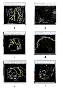
Figure 5. ImageJ Results for Makahiya CAM Assay. Treatment 1 (a) before (b) after; Treatment 2 (c) before (d) after; Treatment 3 (e) before (f) after
The case of makahiya, situated within a specific plant family known for containing phytochemicals with anti-angiogenic properties, serves to reinforce Moerman’s theory of non-random plant selection. This assertion finds further validation in a study by Kutal et al. (2021), which extensively documented the presence of these bioactive compounds within the literature. Such findings suggest a tendency for certain plant families to harbor bioactive compounds with distinct medicinal properties. In line with this, a study conducted by Masagca et al. (2020) investigated the anti-angiogenic effects of makahiya. Their research demonstrated the efficacy of the aqueous root extract of makahiya in suppressing angiogenesis. Additionally, their findings indicated the potentially toxic effects of makahiya extracts, particularly in water and ethanol solutions, on Dalton’s Ascitic Lymphoma cells. These effects may be attributed to mechanisms such as membrane damage, lipid peroxidation, and increased lactate dehydrogenase levels, ultimately leading to a non-apoptotic form of cell death.
In light of Judah Folkman’s theory suggesting that targeting angiogenesis can inhibit tumor growth, the findings suggest that makahiya extract may possess anti-angiogenic properties. In fact, several studies have associated these phytochemicals with potential anti-angiogenic and cancer treatment effects, as supported by Alasvand et al. (2019) and Nasrat et al. (2022). Consequently, the observed effects of makahiya extract underscore its potential as an anti-angiogenic agent with broader implications for cancer treatment strategies.
Efficacy of Different Concentrations of Paper Flower Extract in Alleviating Cell Division
The objective of this study was to evaluate how Paper flower extract alleviates cell division. To achieve this, three distinct doses were administered: treatment 1 (0.1 mL), treatment 2 (0.2 mL), and treatment 3 (0.3 mL). The process involving thirty fertilized duck eggs was chosen as the experimental model, of which nine were for the Paper Flower extract. Following the administration of the Paper Flower extract, which occurred on the 5th day of egg incubation, the treated areas underwent examination using “ImageJ”, to assess the effects of the treatments. The results indicated an anti-angiogenic effect of the extracts, as evidenced by the reduced average diameter of blood vessels compared to the control group, where the average vascularity count is 2.93 at 0.1 mL concentration, while concentrations of 0.2 mL and 0.3 mL showed an average of 5.16 and 1.20, respectively, suggesting a high efficacy rate.
Table 2. Efficacy of Different Concentrations of Paper Flower Extract in Alleviating Cell Division
| Treatments | Vascularity Count | Mean | SD | ||
| R1 | R2 | R3 | |||
| Before Treatment | |||||
| T1 | 9.098 | 12.589 | 12.839 | 11.51 | 2.09 |
| T2 | 15.750 | 13.895 | 10.036 | 13.23 | 2.91 |
| T3 | 10.962 | 13.228 | 7.932 | 10.71 | 2.66 |
| After Treatment | |||||
| T1 | 6.666 | 6.666 | 7.669 | 7.00 | 0.58 |
| T2 | 3.439 | 7.431 | 8.531 | 6.47 | 2.68 |
| T3 | 3.361 | 10.225 | 4.362 | 5.98 | 3.71 |
The statistical analysis in Table 2 confirmed the effectiveness of Paper Flower extracts in inhibiting angiogenesis. Treatment 3 exhibited the highest mean vascularity count (7.00), while treatment 3 showed the lowest (5.98), indicating the highest inhibition of blood vessels. These findings align with the presence of phytochemical compounds found in Paper flowers, making them potential anti-cancer agents due to their capability to inhibit blood vessel growth. Notably, several studies, including those by Kalaiyarasan et al. (2022), Saleem et al. (2019), Teh et al. (2019), and Gupta and Gupta (2021), highlight the promising applications of Paper flowers as natural phytochemical agents.
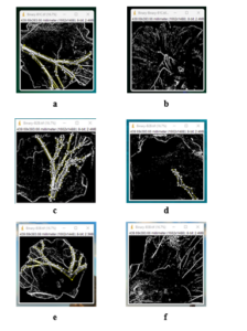
Figure 6. ImageJ Results for Paper flower CAM Assay. Treatment 1 (a) before (b) after; Treatment 2 (c) before (d) after; Treatment 3 (e) before (f) after
Furthermore, the exploration of the molecular mechanisms of plant extracts and phytochemicals by Soni and Duddukuri (2023) sheds light on their diverse targets in cancer progression. In line with this, Hazafa et al. (2020) emphasize the efficacy of polyphenols, including flavonoids, in predicting and managing cancer. These studies collectively support the notion that the phytochemical compounds found in Paper Flowers likely contribute to their anti-angiogenic and potential anti-cancer effects.
Insights from Li and Mashiach (2021) indicate that angiogenesis has a dual role in the inflammatory response and the growth of new blood vessels. This aligns with Folkman’s theory, suggesting that the reduction in blood vessel diameter observed in the statistical analysis could hinder tumor growth. The presence of phytochemical compounds in Paper Flower extract, targeting diverse molecular pathways and inhibiting blood vessel growth, supports Meorman’s theory of synergistic effects, potentially enhancing its therapeutic efficacy against cancer.
Efficacy of Different Concentrations of Makahiya and Paper Flower Extract Mixture in Alleviating Cell Division
The study determined the effectiveness of the mixture extract of makahiya and Paper Flower in alleviating cell division with three different treatments: Treatment 1 – 0.1 mL; Treatment 2 – 0.2 mL; and Treatment 3 – 0.3 mL. A total of nine duck eggs were used in this extraction, three eggs per replication. The effectiveness of the extract mixture was demonstrated through a reduction in vascularity counts after each treatment at different concentrations. In the initial treatment with a concentration of 0.1ml, the mean vascularity count dropped from 12.19 before treatment to 6.27 afterward, indicating a substantial decrease of 5.92 in vascularity. Similarly, treatments using concentrations of 0.2ml and 0.3ml showed notable reductions in vascularity counts, with decreases from 11.65 to 3.79, a difference of 7.86, and from 11.63 to 4.30, a difference of 7.33, respectively. These results highlighted the extract’s effectiveness in reducing vascularity, as evidenced by the notable differences observed between the before and after treatment counts across the different concentrations.
Table 3. Efficacy of Different Concentrations of Makahiya and Paper Flower Extract Mixture in Alleviating Cell Division
| Treatments | Vascularity Count | Mean | SD | ||
| R1 | R2 | R3 | |||
| Before Treatment | |||||
| T1 | 14.103 | 12.192 | 10.283 | 12.19 | 1.91 |
| T2 | 13.641 | 11.724 | 9.591 | 11.65 | 2.03 |
| T3 | 12.781 | 12.308 | 9.802 | 11.63 | 1.60 |
| After Treatment | |||||
| T1 | 6.343 | 7.286 | 5.193 | 6.27 | 1.05 |
| T2 | 6.952 | 1.369 | 3.051 | 3.79 | 2.86 |
| T3 | 7.263 | 0.520 | 5.116 | 4.30 | 3.44 |
In this table, prior to the treatment, T1 exhibited the highest vascularity count, along with the highest mean of 12.19. Subsequently, following treatment involving the administration of the mixed extraction of makahiya and Paper flower, a reduction in this value was observed, with the mean decreasing to 6.27. Notably, among the treatments, T2 demonstrated the most pronounced effect of the extraction, as evidenced by its mean decreasing substantially from 11.65 to 3.79. These results align with findings from Mondal et al. (2019) and Hanggag and Elhaw (2022), which also indicate the presence of anti-cancer properties in the makahiya extract and Paper flower. Such properties likely contribute to the observed inhibition of blood vessels.
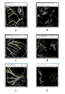
Figure 7. ImageJ Results for Makahiya and Paper Flower CAM Assay. Treatment 1 (a) before (b) after; Treatment 2 (c) before (d) after; Treatment 3 (e) before (f) after
Collectively, these studies suggest that phytochemicals hold promising implications for anti-angiogenic capabilities, thus presenting a potential avenue for further exploration in cancer treatment strategies. Moreover, Javed (2020) underscores the significance of medicinal plants as abundant sources of compounds like artemisinin, vinblastine, and vincristine, which have made contributions to drug development, further emphasizing the importance of continued research in harnessing the therapeutic potential of phytochemicals for combating cancer in both plants.
Significant Difference in the Effectiveness of Different Concentrations of Makahiya and Paper Flower Extract in Alleviating Cell Division
Table 4 shows the results of a one-way analysis of variance to determine the significance of the difference in the vascularity count in different treatment groups before the treatment. It can be observed that the F value is 0.899 with 9 and 20 degrees of freedom. The p-value is 0.544 which is greater than 0.05. This entails failure of rejecting the null hypothesis, indicating that there is no significant difference in the vascularity count of samples in different treatment groups. Further, this means that all samples in different treatment groups are comparable in terms of vascularity count before or at the start of the experiment.
Table 4. Significant Difference in the Vascularity Count in Different Treatment Groups Before Treatment
| Sum of Squares | df | Mean Square | F | p | Decision | |
| Between Groups | 44.28 | 9 | 4.92 | 0.899 | 0.544 | Not Significant |
| Within Groups | 109.44 | 20 | 5.47 | |||
| Total | 153.72 | 29 |
Moreover, table 5 shows the results of a one-way analysis of variance to determine the significance of the difference in the effectiveness of different concentrations of makahiya and Paper flower extract in alleviating cell division in terms of vascularity count after treatment. It can be observed that the F value is 5.478 with 9 and 20 degrees of freedom. The p-value is 0.001, which is less than 0.05. This further means that the null hypothesis should be rejected, indicating that at least one among the ten treatments significantly differs from the other in terms of its effectiveness in alleviating cell division.
Table 5. Significant Difference in the Effectiveness of Different Concentrations of Makahiya and Paper flower Extract in terms of Vascularity Count After Treatment
| Sum of Squares | df | Mean Square | F | p | Decision | |
| Between Groups | 633.53 | 9 | 70.392 | 5.478 | 0.001 | Reject H0 |
| Within Groups | 257.02 | 20 | 12.85 | (Significant) | ||
| Total | 890.55 | 29 |
To determine which among the treatments significantly differ from the other, post hoc analysis was conducted particularly the pair-wise comparisons of sample means via the Tukey HSD test. The Tukey’s honestly significant difference test (Tukey’s HSD) is used to test differences among sample means for significance. The Tukey’s HSD tests all pairwise differences while controlling the probability of making one or more Type I errors. Tukey’s Honestly Significant Difference test (Tukey’s HSD) is widely recognized and frequently employed as a prominent technique for minimizing the error rate, thereby facilitating the derivation of precise and meaningful conclusions (Nanda et al., 2021).
Meanwhile, Table 6 shows the values of the control group which has three replications, therefore the vascularity count of the replications when the egg was first opened and after 24 hours, was added and was divided into three to obtain the values of the mean. Thus, the mean of the replications before the 24-hour count was valued at 11.010, while the measurement after 24 hours was 19.015, which implied that the measurement of the blood vessels notably increased after 24 hours with zero treatments added. This is in line with the study of Gupta et al. (2022), where the eggs utilized for the control group were left untreated up until the evaluation of the experimental groups.
Table 6. Control Group Measurements
| Vascularity Count | ||||
| R1 | R2 | R3 | Mean | |
| Before 24 hours | 9.731 | 10.954 | 12.345 | 11.010 |
| After 24 hours | 9.731 | 22.946 | 24.367 | 19.015 |
Table 7 shows the results of the post hoc comparisons using the Tukey HSD test. This test was done to determine which of the four treatments have significant differences in terms of effectiveness. Since there are 10 treatments, the researchers decided to report only those comparisons with significant differences. It can be observed that significant differences occur between the alternative treatments and the control treatment, as indicated by the p-values which are less than 0.05. The results also show a negative mean difference between these treatments, indicating that for each pair, the control treatment is significantly more effective than the alternative treatment in alleviating cell division.
Table 7. Post Hoc Comparisons using the Tukey HSD Test
| Mean
Difference |
p | Decision | Interpretation | |
| Between T1 and Control | -16.087 | 0.001 | Reject H0 | Significant Difference |
| Between T2 and Control | -13.855 | 0.004 | Reject H0 | Significant Difference |
| Between T3 and Control | -17.813 | 0.000 | Reject H0 | Significant Difference |
| Between T4 and Control | -12.014 | 0.015 | Reject H0 | Significant Difference |
| Between T5 and Control | -12.548 | 0.010 | Reject H0 | Significant Difference |
| Between T6 and Control | -13.032 | 0.007 | Reject H0 | Significant Difference |
| Between T7 and Control | -12.740 | 0.009 | Reject H0 | Significant Difference |
| Between T8 and Control | -15.224 | 0.001 | Reject H0 | Significant Difference |
| Between T9 and Control | -14.715 | 0.002 | Reject H0 | Significant Difference |
Overall, the three extracts exhibited notable differences in terms of their effectiveness in alleviating cell division, with the makahiya extract with the 0.3 mL plant concentration demonstrating the highest anti-angiogenesis efficacy, followed by the combined extract of makahiya and Paper flower, and lastly, the Paper flower extract. The result agrees with the statement of Chojnacka and Lewandowska (2019) in which the polyphenols abundant in makahiya impede angiogenesis by impacting vital factors and cellular processes, restricting the formation of blood vessels, and limiting the migration and invasion of cancer cells, thereby leading to a reduction in tumor growth. Moreover, Ribeiro et al. (2018) expounded that natural phytochemicals from plants or micro fungi show significant potential in treating angiogenesis-related conditions with lower toxicity than synthetics. This directed attention towards exploring the effectiveness of natural compounds as angiogenesis modulators, offering new therapies with reduced drug reliance and associated side effects.
Furthermore, these results show the potential of the different plant phytochemicals in terms of their antiangiogenic attributes due to the noticeable reduction in vascularity counts prior to and after the different treatments, which reflected the studies of Bole et al. (2021), Kumar (2020), and Rizwan et al. (2022), which identified the presence of 40 chemical components, including alkaloids, phenols, and flavonoids, for the makahiya plant. Additionally, in the study relating to the paper flower, Gupta and Gupta (2021) state that paper flowers were found to contain a variety of substances, including fat, tannin, glycosides, anthraquinone, terpenoids, and saponin. Alkaloids, flavonoids, phenolic compounds, and tannins were identified by the use of specific qualitative assays designed to identify these particular phytochemicals. These phytochemicals, such as alkaloids and flavonoids, have anticancer properties, as mentioned in several studies by Mondal et al. (2019), Kurek (2019), Bisol et al. (2019), George et al. (2021), and Gurler (2020).
Conclusion
Cancer is a widespread illness that has a long-term impact on patients from various countries. There is a scarcity of safer methods to hinder the growth of tumor cells compared to the existing treatments like radiotherapy and chemotherapy, and there is a lack of utilizing naturally occurring anticancer properties. Hence, this study investigated the efficacy of naturally occurring anticancer properties in inhibiting tumor angiogenesis through makahiya and Paper flower extracts. The results demonstrated that the makahiya extract, particularly at a 0.3 mL plant concentration, exhibited the highest effectiveness in alleviating cell division with the lowest post-treatment vascularity mean of 1.20. This was followed by the combined extract of makahiya and Paper flower at 0.2 mL, which showed a post-treatment vascularity mean of 3.79, and lastly, the Paper flower extract at 0.2 mL, which resulted in a post-treatment vascularity mean of 6.46.
Recommendation
In light of the study’s findings, the researchers recommend exploring more innovative techniques to improve the accuracy of CAM assay angiogenesis studies. Furthermore, future researchers are advised to extend the duration of CAM assays for thorough evaluation and to consider alternative test subjects to avoid adverse effects, such as those observed with the Paper flower extract. Future studies should examine the toxicity of certain compounds that may cause cellular damage, ensuring the safety and effectiveness of plant-based applications. Conducting toxicity studies is essential before any potential human or animal applications, and the targeted application of extracts to specific inhibition areas is recommended to enhance effectiveness. Finally, the researchers call for the exploration of new methods and tools for accurately analyzing blood vessel growth beyond the current ImageJ software.